


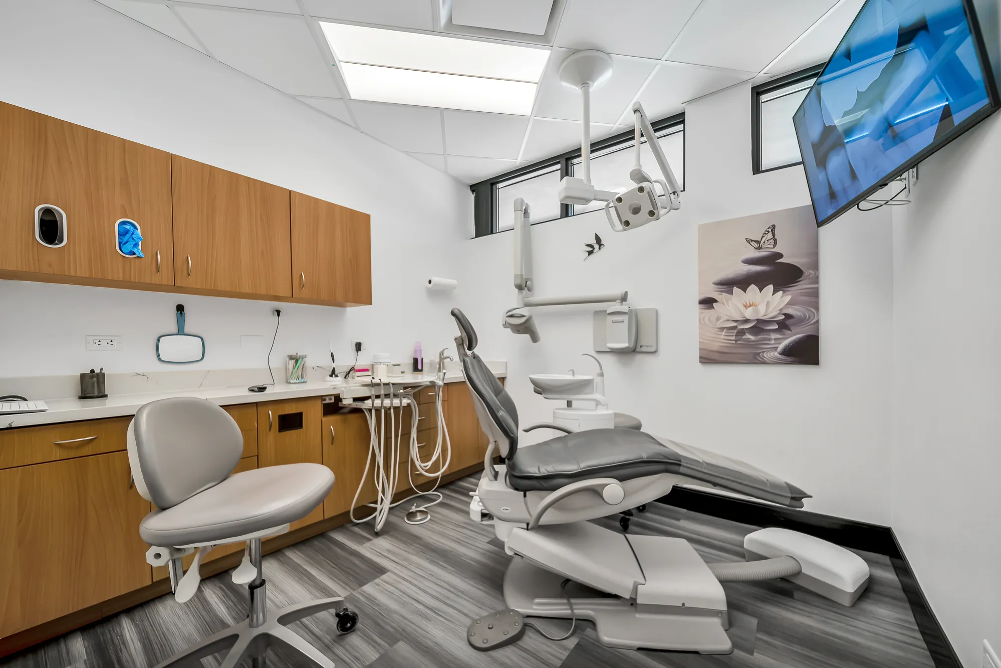
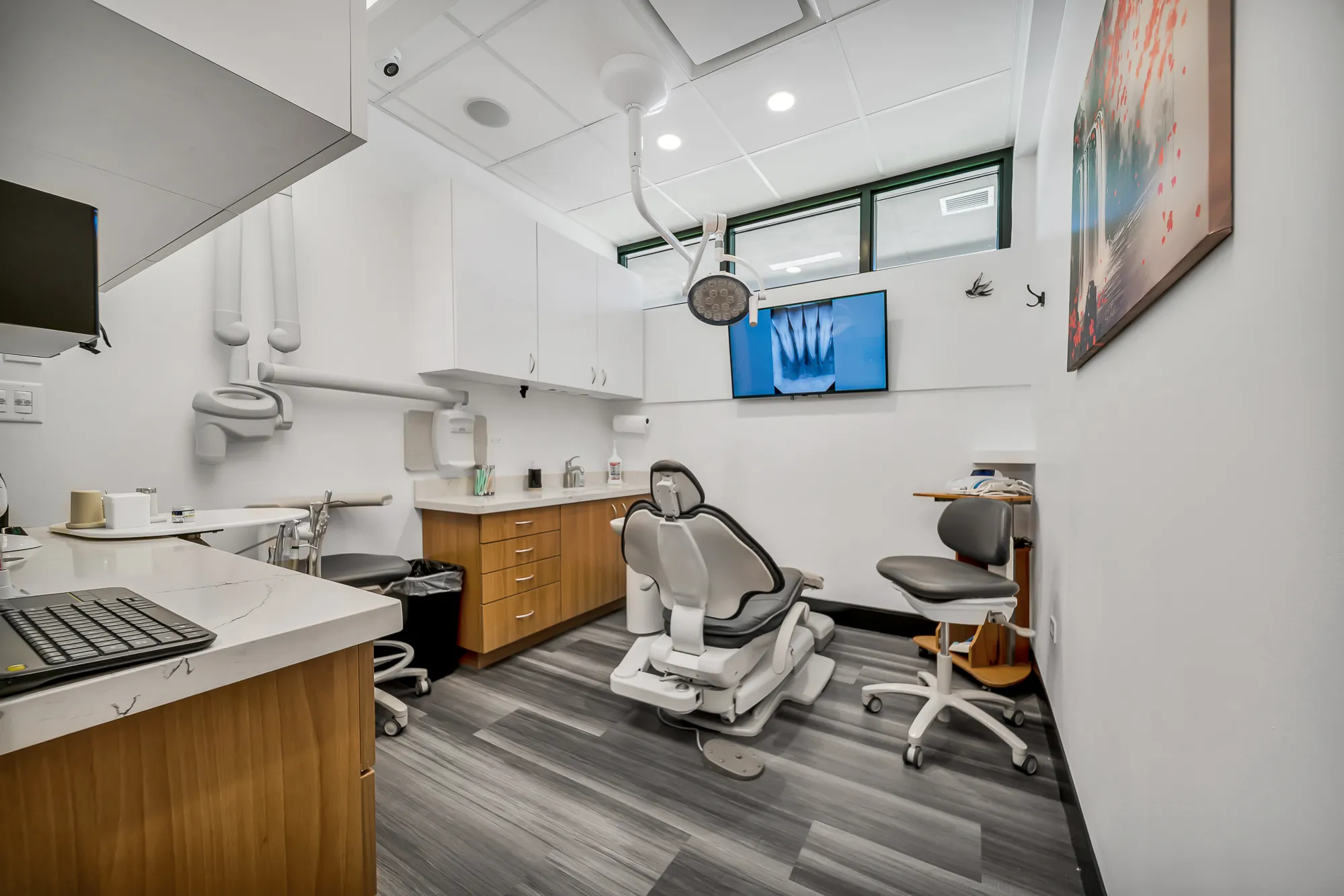


3D Imaging for Dental Implants
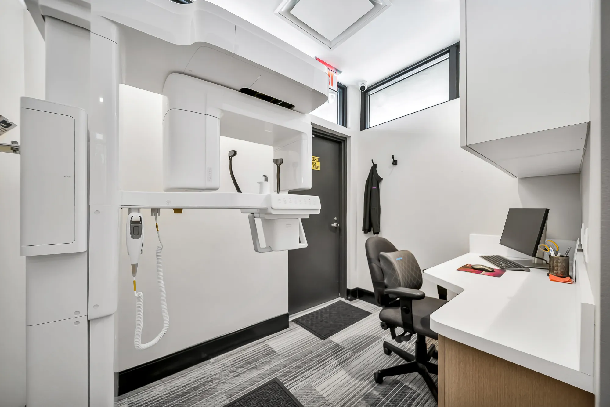
Dental cone beam computed tomography (CT) is a special type of x-ray equipment used when regular dental or facial x-rays are not sufficient. Dr. Chakalov may use this technology to produce three dimensional (3-D) images of your teeth, soft tissues, nerve pathways and bone in a single scan. This procedure requires little to no special preparation. Tell us if there is a possibility you are pregnant. Wear loose, comfortable clothing and leave jewelry at home. You may be asked to wear a gown.
What are some common uses of the procedure?
Dental cone beam CT is commonly used for treatment planning of cases involving:
- accurate planning and placement of dental implants.
- surgical planning for removal of impacted teeth.
- determining bone structure and tooth orientation.
- evaluation of the jaw, sinuses, nerve canals and nasal cavity.
- detecting, measuring and treating jaw tumors.
- diagnosing temporomandibular joint disorder (TMJ).
- locating the origin of pain or pathology.
- cephalometric analysis.
- reconstructive surgery.
What are the benefits vs. risks?
Benefits:
- Cone beam CT scans provide more information that conventional dental x-ray, allowing for more precise treatment planning.
- A single scan produces a wide variety of views and angles that can be manipulated to provide a more complete evaluation.
- The focused x-ray beam reduces scatter radiation, resulting in better image quality.
- CT scanning is painless, noninvasive and accurate.
- A major advantage of CT is its ability to image bone and soft tissue at the same time.
- No radiation remains in a patient’s body after a CT examination.
- X-rays used in CT scans should have no immediate side effects.
Risks:
- There is always a slight chance of cancer from excessive exposure to radiation.
- However, the benefit of an accurate diagnosis far outweighs the risk.
- CT scanning is, in general, not recommended for pregnant women unless medically necessary because of potential risk to the baby in the womb.